
CT & MRI Scans
CT and MRI scans are advanced diagnostic tools offered at Dr. Ankur Agrawal’s Sports Injury Center in Bareilly, providing detailed imaging of bones, joints, muscles, and soft tissues to accurately diagnose complex orthopedic and sports-related conditions. These scans go beyond standard X-rays, helping detect issues that may not be visible otherwise.
A CT (Computed Tomography) scan uses multiple X-ray images combined with computer processing to create cross-sectional views of bones and joints. It is especially useful in identifying subtle fractures, bone tumors, joint misalignments, and evaluating joint structures in 3D. Dr. Agrawal often recommends CT scans for patients with traumatic injuries or complex bone problems where precision is critical.
An MRI (Magnetic Resonance Imaging) provides even more detailed images of soft tissues such as ligaments, tendons, cartilage, and spinal discs. It uses magnetic fields and radio waves instead of radiation, making it safe and non-invasive. MRI is ideal for diagnosing ligament tears (like ACL injuries), meniscus damage, herniated discs, and chronic joint pain with unclear causes.
✅ What is a CT Scan?
A CT (Computed Tomography) scan uses X-rays and a computer to create detailed cross-sectional images of the body. It shows bones, organs, blood vessels, and soft tissues more clearly than regular X-rays.
✅ How Does CT Work?
The patient lies on a table that slides into a circular machine.
X-rays are rotated around the body.
A computer assembles the data into detailed images.
It is quick (usually takes 5–10 minutes).
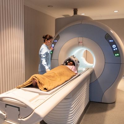
✅ What is an MRI?
An MRI (Magnetic Resonance Imaging) uses strong magnets and radio waves to create high-resolution images of soft tissues like the brain, spinal cord, muscles, joints, and organs.
✅ How Does MRI Work?
The patient lies in a tube-like scanner.
Magnetic fields align hydrogen atoms in the body.
Radio waves disrupt the atoms briefly, then signals are sent to a computer.
These signals are used to build detailed images of internal structures.
MRI takes longer (20–45 minutes) and does not use radiation.
✅ When is a CT Scan Needed?
A CT scan is recommended when:
There’s a suspected bone fracture, especially in complex areas (spine, pelvis, skull)
After accidents to check for internal bleeding or organ damage
To evaluate chest, lungs, or abdominal issues
For detecting tumors, blood clots, infections, or kidney stones
During emergency situations needing fast and clear images
✅ When is an MRI Needed?
An MRI is used when:
Detailed images of soft tissues are needed (brain, spinal cord, muscles, joints)
There’s chronic joint pain, nerve injury, or disc problem
To detect brain tumors, multiple sclerosis, or strokes
For sports injuries involving ligaments, cartilage, or tendons
To examine internal organs like liver, uterus, or prostate in more detail